Pelvic Anatomy Bones - Pelvis Problems Johns Hopkins Medicine / Many muscles that move the trunk and legs, such as our abdominal muscles, attach to the hip bones.. Its symphyseal surface unites with the pubis of the opposite side to form the pubic symphysis; Search other sites for 'pelvic bone' nlm pubmed google websites google images quackwatch drugstore.com. In addition, the broad hip bones provide protection to the delicate internal organs of the pelvis, such as the intestines, urinary bladder, and uterus. The outlet is formed by the pubic arch, ischial spines, sacrotuberous ligaments, and the coccyx. There are two hip bones, one on the left side of the body and the other on the right.
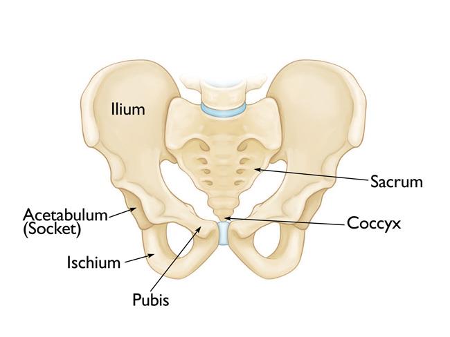
The bones of the pelvis are the hip bones, sacrum, and coccyx. These two bones approach each other at the front and form a joint called the pubic symphysis. The pelvis consists of the sacrum, the coccyx, the ischium, the ilium, and the pubis. The ilium, pubis and ischium. The pelvis is an anatomically complex and functionally informative bone that contributes directly to both human locomotion and obstetrics.
Wesnorman Com Anatomy Lateral View Of The Hip Pelvis Pelvis Anatomy Medical Anatomy Human Anatomy And Physiology from i.pinimg.com Hip skeleton on blue background. Together, they form the part of the pelvis called the pelvic girdle. As well as the coccyx, a small tail like bone attaching to the underside of the sacrum. The counterrotation of the bones continues all down the leg and through the foot. Vector illustration isolated on a white background. The pelvic bones and the sacrum. Many muscles that move the trunk and legs, such as our abdominal muscles, attach to the hip bones. There are two hip bones, one on the left side of the body and the other on the right.
In this video we discuss the bones of the hip, the structure of the hip and the anatomy of the pelvic girdle.
Search other sites for 'pelvic bone' nlm pubmed google websites google images quackwatch drugstore.com. A 3d rotatable model of the bony structures of the pelvis: The pelvis consists of two innominate bones and the sacrum to which coccyx is attached. The low back is defined by the lumbar spine, and the pelvis is defined by the bones of the pelvic girdle. The hip bones join to the upper part of. The hip bone has three parts: The pelvic spine is the posterior portion of the pelvis below the lumbar spine, composed of the sacrum and coccyx. The inlet to the pelvic canal is at the level of the sacral promontory and superior aspect of the pubic bones. The bones of the pelvis are the hip bones, sacrum, and coccyx. The pelvis is a ring of bones located at the lower end of the trunk—between the spine and the legs. The outlet is formed by the pubic arch, ischial spines, sacrotuberous ligaments, and the coccyx. When you are taking anatomy and physiology you will be required to know the anatomical structure locations of the pelvis. Human pelvis image human male anatomy scheme.
The counterrotation of the bones continues all down the leg and through the foot. During childhood, these sections are separate bones, joined by the triradiate cartilage. General anatomy of the pelvis. The right and left hip bones, sacrum, and coccyx. The pelvis is a boney structure at the base of the lumbar spine.
Pelvic Fractures Orthoinfo Aaos from orthoinfo.aaos.org The pelvis consists of the sacrum, the coccyx, the ischium, the ilium, and the pubis. The ilium, ischium and the pubic bone. There are four articulations within the pelvis: Each hip bone contains three bones — the ilium, ischium, and pubis — that fuse together as we grow older. Learning pelvic anatomy is composed of learning bones, muscles, ligaments, nerves and vascular supply. Hip skeleton on blue background. The pelvis helps anchor the muscles and protect the organs in the lower abdomen. The right and left hip bones, sacrum, and coccyx.
The right and left hip bones, sacrum, and coccyx.
The pelvic bones include the: The complex intersection of pelvic … Its symphyseal surface unites with the pubis of the opposite side to form the pubic symphysis; Search other sites for 'pelvic bone' nlm pubmed google websites google images quackwatch drugstore.com. The pelvic girdle (hip girdle) is formed by a single bone, the hip bone or coxal bone (coxal = hip), which serves as the attachment point for each lower limb. There are two hip bones, one on the left side of the body and the other on the right. During childhood, these sections are separate bones, joined by the triradiate cartilage. The right and left hip bones, sacrum, and coccyx. Trauma in pregnancy shoulder dystocia management hip anatomy femur bone male pelvis sacroiliac dysfunction pubic symphysis pediatric limp pelvic fracture ischiofemoral impingement. The counterrotation of the bones continues all down the leg and through the foot. These two bones approach each other at the front and form a joint called the pubic symphysis. In humans they are triangular and lie on the upper back between the levels of the second and eighth ribs. In addition, the broad hip bones provide protection to the delicate internal organs of the pelvis, such as the intestines, urinary bladder, and uterus.
Vector illustration isolated on a white background. A 3d rotatable model of the bony structures of the pelvis: During childhood, these sections are separate bones, joined by the triradiate cartilage. Because of the pelvis' important role in obstetrics, it is one of the most sexually dimorphic bony elements of the human body. It is based on a 3d scan.
Bones Of The Lumbar Spine And Pelvis from www.learnmuscles.com Sacrum (the large triangular bone at the base of the spine) coccyx (tailbone) hip bones; It is composed of four major bones: Each pelvic bone (hip bone) is made by the combination three bones namely, the ilium, pubis, and ischium. Human pelvis image human male anatomy scheme. As well as the coccyx, a small tail like bone attaching to the underside of the sacrum. Learning pelvic anatomy is composed of learning bones, muscles, ligaments, nerves and vascular supply. When you are taking anatomy and physiology you will be required to know the anatomical structure locations of the pelvis. A full understanding of pelvic anatomy is required to treat pelvic fractures, to prevent iatrogenic injuries, and to provide the best results.
Its body forms 1/5 of the acetabulum;
The low back is defined by the lumbar spine, and the pelvis is defined by the bones of the pelvic girdle. The pelvis is an anatomically complex and functionally informative bone that contributes directly to both human locomotion and obstetrics. The pelvic skeleton is formed posteriorly (in the area of the back), by the sacrum and the coccyx and laterally and anteriorly (forward and to the sides), by a pair of hip bones. The pelvis consists of two innominate bones and the sacrum to which coccyx is attached. It is based on a 3d scan. The pelvic bones and the sacrum. Each innominate bone includes three other bones named ilium, ischium, and pubis. The hip bone has three parts: It is composed of four major bones: As well as the coccyx, a small tail like bone attaching to the underside of the sacrum. Its symphyseal surface unites with the pubis of the opposite side to form the pubic symphysis; The bones of the pelvis are the hip bones, sacrum, and coccyx. These two bones approach each other at the front and form a joint called the pubic symphysis.
Trauma in pregnancy shoulder dystocia management hip anatomy femur bone male pelvis sacroiliac dysfunction pubic symphysis pediatric limp pelvic fracture ischiofemoral impingement pelvic anatomy. Learning pelvic anatomy is composed of learning bones, muscles, ligaments, nerves and vascular supply.
Pelvic Anatomy Bones - Pelvis Problems Johns Hopkins Medicine / Many muscles that move the trunk and legs, such as our abdominal muscles, attach to the hip bones.. There are any Pelvic Anatomy Bones - Pelvis Problems Johns Hopkins Medicine / Many muscles that move the trunk and legs, such as our abdominal muscles, attach to the hip bones. in here.